Anatomy Of Chest / Human Thorax Anatomy. Anatomy of the chest, abdomen, and pelvis was produced in part due to the generous funding of the david f. Stability to arm and shoulder movement; Anatomy articles covering gross anatomy, microscopic anatomy, natural variants, and pathophysiologic variants with accompanying images. Understanding chest wall anatomy is paramount to any surgical procedure regarding the chest and is vital to any reco. In vertebrates (fishes, amphibians, reptiles, birds, and mammals), the thorax is the chest, with the chest being that part of the body between the neck and the abdomen.
Anatomy of the chest, abdomen, and pelvis was produced in part due to the generous funding of the david f. The visceral pleura invests the lungs. And flexibility to aid in the functional process of respiration. Anatomy articles covering gross anatomy, microscopic anatomy, natural variants, and pathophysiologic variants with accompanying images. Other important structures, such as the pleura, only become visible when abnormal, and some are not visible at all, such as the phrenic nerve.
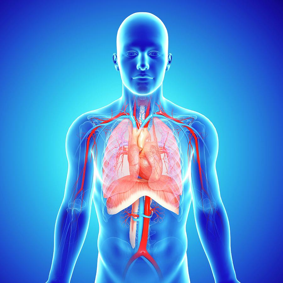
Chest Anatomy Photograph by Pixologicstudio/science Photo Library from images.fineartamerica.com And flexibility to aid in the functional process of respiration. An awareness of the range of normal is important, but the best tip is to look for increase in density as well as size. The visceral pleura invests the lungs. Jul 07, 2016 · the chest wall is a complex system that provides rigid protection to the vital organs such as the heart, lungs, and liver; In vertebrates (fishes, amphibians, reptiles, birds, and mammals), the thorax is the chest, with the chest being that part of the body between the neck and the abdomen. Jul 16, 2019 · in addition to moving the arm and pectoral girdle, muscles of the chest and upper back work together as a group to support the vital process of breathing. Oct 15, 2017 · radiology basics of chest ct anatomy with annotated coronal images and scrollable axial images to help medical students and junior doctors learning anatomy. Other important structures, such as the pleura, only become visible when abnormal, and some are not visible at all, such as the phrenic nerve.
Stability to arm and shoulder movement;
Jul 07, 2016 · the chest wall is a complex system that provides rigid protection to the vital organs such as the heart, lungs, and liver; Jul 16, 2019 · in addition to moving the arm and pectoral girdle, muscles of the chest and upper back work together as a group to support the vital process of breathing. The visceral pleura invests the lungs. Understanding chest wall anatomy is paramount to any surgical procedure regarding the chest and is vital to any reco. Thorax, the part of an animal's body between its head and its midsection. Other important structures, such as the pleura, only become visible when abnormal, and some are not visible at all, such as the phrenic nerve. Stability to arm and shoulder movement; An awareness of the range of normal is important, but the best tip is to look for increase in density as well as size. In addition, important neurovascular bundles course along each rib, containing an intercostal nerve, artery, and vein. Oct 15, 2017 · radiology basics of chest ct anatomy with annotated coronal images and scrollable axial images to help medical students and junior doctors learning anatomy. Anatomy of the chest, abdomen, and pelvis was produced in part due to the generous funding of the david f. The hila are often wrongly called abnormal when normal and normal when abnormal. And flexibility to aid in the functional process of respiration.
The vertebrate thorax contains the chief organs of In vertebrates (fishes, amphibians, reptiles, birds, and mammals), the thorax is the chest, with the chest being that part of the body between the neck and the abdomen. Anatomy articles covering gross anatomy, microscopic anatomy, natural variants, and pathophysiologic variants with accompanying images. Oct 15, 2017 · radiology basics of chest ct anatomy with annotated coronal images and scrollable axial images to help medical students and junior doctors learning anatomy. Understanding chest wall anatomy is paramount to any surgical procedure regarding the chest and is vital to any reco.
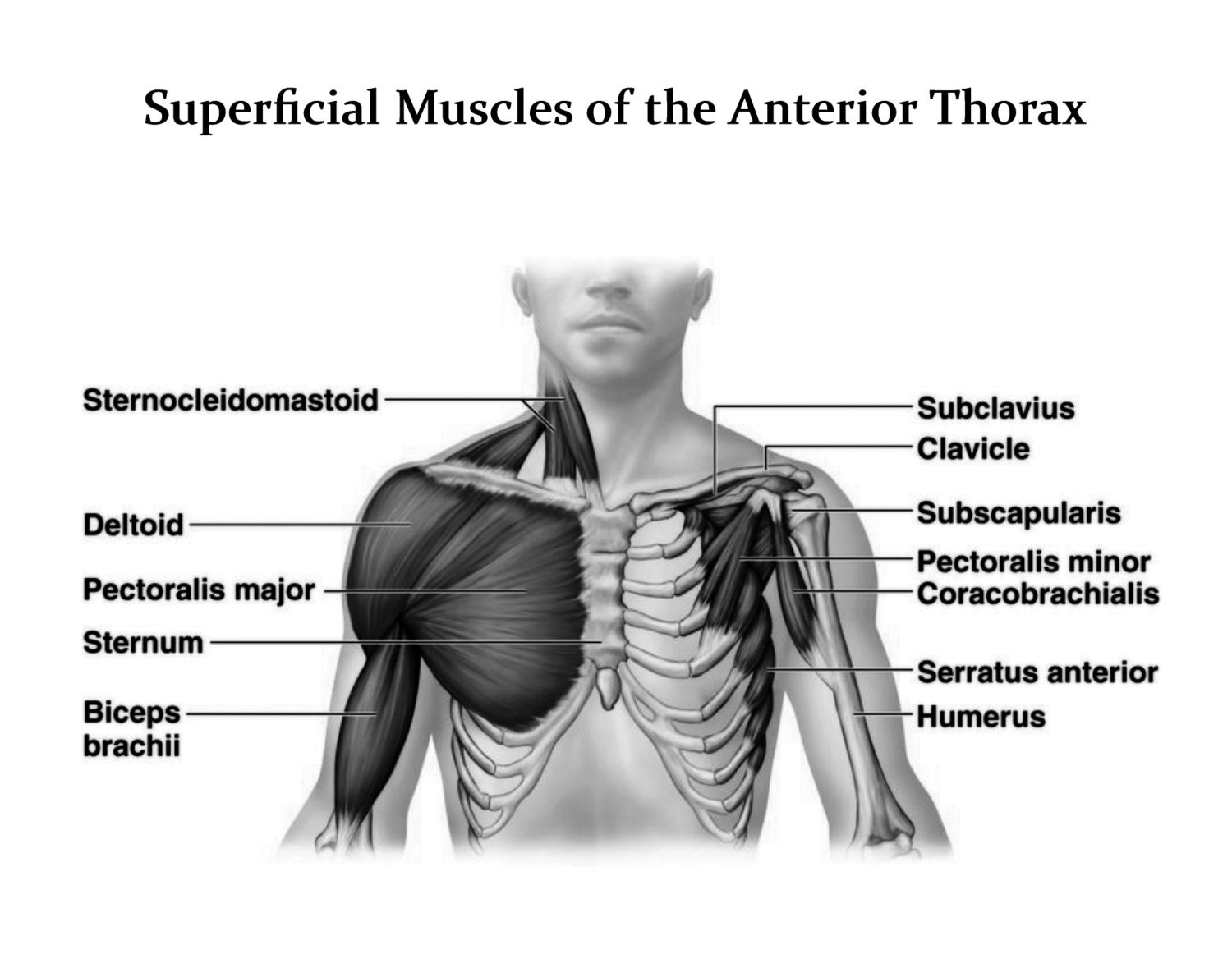
Chest Muscle Anatomy Diagram / Chest Muscles Anatomy Chest Muscles Anatomy - Anatomy ... - More ... from img0.etsystatic.com Stability to arm and shoulder movement; Understanding chest wall anatomy is paramount to any surgical procedure regarding the chest and is vital to any reco. The vertebrate thorax contains the chief organs of An awareness of the range of normal is important, but the best tip is to look for increase in density as well as size. And flexibility to aid in the functional process of respiration. The inner lining of the chest wall is the parietal pleura. The hila are often wrongly called abnormal when normal and normal when abnormal. Thorax, the part of an animal's body between its head and its midsection.
The vertebrate thorax contains the chief organs of
Oct 15, 2017 · radiology basics of chest ct anatomy with annotated coronal images and scrollable axial images to help medical students and junior doctors learning anatomy. The inner lining of the chest wall is the parietal pleura. The vertebrate thorax contains the chief organs of In vertebrates (fishes, amphibians, reptiles, birds, and mammals), the thorax is the chest, with the chest being that part of the body between the neck and the abdomen. Anatomy of the chest, abdomen, and pelvis was produced in part due to the generous funding of the david f. Sep 22, 2020 · the chest wall is composed of layers of muscle, bony ribs, costal cartilages, sternum, clavicles, and scapulae. And flexibility to aid in the functional process of respiration. Other important structures, such as the pleura, only become visible when abnormal, and some are not visible at all, such as the phrenic nerve. Swensen fund for innovation in teaching. The hila are often wrongly called abnormal when normal and normal when abnormal. Anatomy articles covering gross anatomy, microscopic anatomy, natural variants, and pathophysiologic variants with accompanying images. An awareness of the range of normal is important, but the best tip is to look for increase in density as well as size. Jul 07, 2016 · the chest wall is a complex system that provides rigid protection to the vital organs such as the heart, lungs, and liver;
The visceral pleura invests the lungs. The vertebrate thorax contains the chief organs of In vertebrates (fishes, amphibians, reptiles, birds, and mammals), the thorax is the chest, with the chest being that part of the body between the neck and the abdomen. Stability to arm and shoulder movement; The inner lining of the chest wall is the parietal pleura.
Human chest anatomy, illustration - Stock Image - F011/5850 - Science Photo Library from media.sciencephoto.com Stability to arm and shoulder movement; Jul 07, 2016 · the chest wall is a complex system that provides rigid protection to the vital organs such as the heart, lungs, and liver; Other important structures, such as the pleura, only become visible when abnormal, and some are not visible at all, such as the phrenic nerve. The inner lining of the chest wall is the parietal pleura. In vertebrates (fishes, amphibians, reptiles, birds, and mammals), the thorax is the chest, with the chest being that part of the body between the neck and the abdomen. And flexibility to aid in the functional process of respiration. The visceral pleura invests the lungs. An awareness of the range of normal is important, but the best tip is to look for increase in density as well as size.
Anatomy articles covering gross anatomy, microscopic anatomy, natural variants, and pathophysiologic variants with accompanying images.
Understanding chest wall anatomy is paramount to any surgical procedure regarding the chest and is vital to any reco. In addition, important neurovascular bundles course along each rib, containing an intercostal nerve, artery, and vein. Jul 16, 2019 · in addition to moving the arm and pectoral girdle, muscles of the chest and upper back work together as a group to support the vital process of breathing. Oct 15, 2017 · radiology basics of chest ct anatomy with annotated coronal images and scrollable axial images to help medical students and junior doctors learning anatomy. The vertebrate thorax contains the chief organs of The visceral pleura invests the lungs. An awareness of the range of normal is important, but the best tip is to look for increase in density as well as size. Stability to arm and shoulder movement; The hila are often wrongly called abnormal when normal and normal when abnormal. Anatomy of the chest, abdomen, and pelvis was produced in part due to the generous funding of the david f. The inner lining of the chest wall is the parietal pleura. Other important structures, such as the pleura, only become visible when abnormal, and some are not visible at all, such as the phrenic nerve. In vertebrates (fishes, amphibians, reptiles, birds, and mammals), the thorax is the chest, with the chest being that part of the body between the neck and the abdomen.